Dental Implants In Worcester: A Patient Guide to Dental Implants
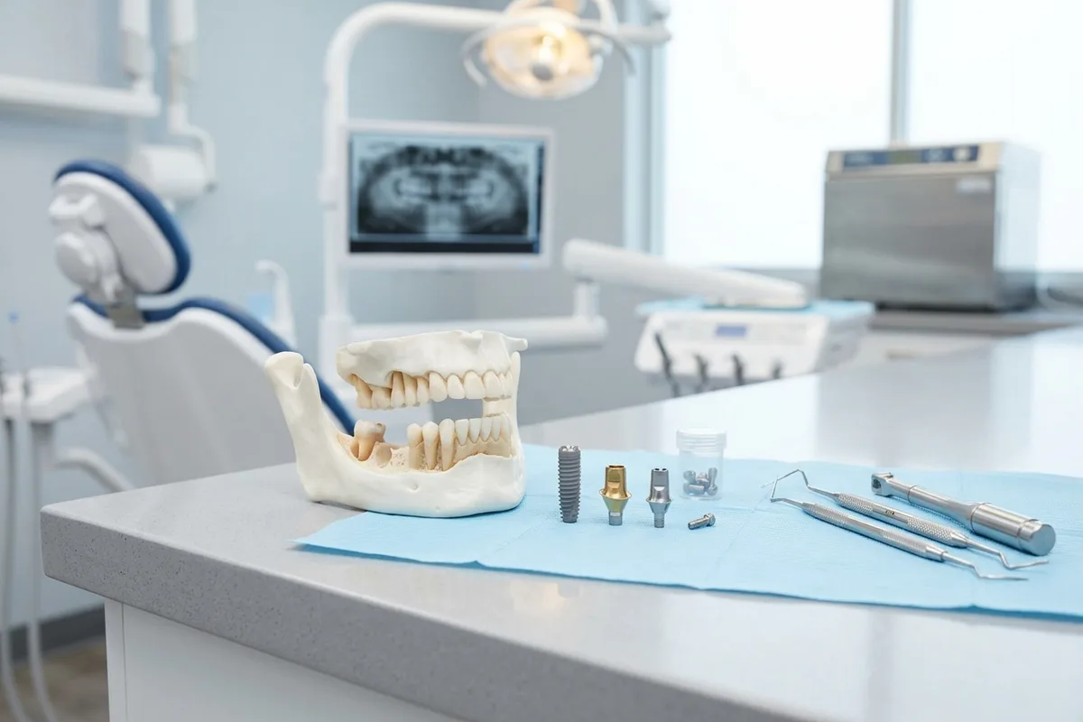
When evaluating dental implants in Worcester, understanding clinical candidacy, surgical steps, and long-term hygiene helps patients navigate restorative options. This guide outlines how dental fixtures integrate with bone to support natural oral function.
Short Answer
Short Answer
Receiving dental implants in Worcester involves surgically anchoring biocompatible posts into the jaw to permanently replace missing teeth. This multi-phase treatment requires sufficient bone density, healthy gums, and a thorough anatomical evaluation. Once the titanium or ceramic fixture biologically bonds to the bone, a custom crown is attached, providing stable, long-lasting chewing ability and structural stability for surrounding oral tissues.[1][2]
Understanding dental implants and local care options
Modern dental implant therapy replaces lost tooth roots with biocompatible surgical fixtures anchored directly within the jawbone. When exploring local treatment paths, patients find that successful implant placement depends on careful preoperative planning, healthy local tissues, and consistent communication with a restorative dental provider throughout the entire multi-stage process.[1][2]
Defining endosseous implant therapy
An endosseous implant is a medical device surgically embedded directly into the alveolar bone of the maxilla or mandible. Unlike traditional removable prosthetics, these fixtures act as artificial roots, stimulating bone and holding fixed crowns, bridges, or overdentures securely in place to restore masticatory function and normal speech.[1][2]
The role of localized ongoing care
Undergoing care with a nearby dental team ensures continuous surveillance across each phase of healing and restoration. Because successful integration requires structured follow-up visits, having accessible care simplifies routine surgical site assessments, suture removal, healing monitoring, and immediate coordination between the surgical team and the restorative practitioner.[1][2]
Primary factors influencing implant candidacy
Candidacy for dental implant placement depends heavily on individual anatomical stability and general health factors. Dentists evaluate both oral conditions and systemic medical histories to ensure a patient can safely undergo surgery and achieve predictable, long-term biological integration between the bone and the implant fixture.[1][2]
Bone morphology and volume requirements
Sufficient bone height, width, and density are fundamental requirements for establishing primary implant stability. When teeth have been missing for an extended period, the surrounding alveolar bone naturally resorbs. In such instances, patients may require supplemental bone grafting or sinus augmentation to build adequate bone volume before implant placement.[1][2]
Systemic health conditions and habits
Systemic issues like poorly managed diabetes or active autoimmune conditions can slow tissue recovery and impair bone fusion. Furthermore, tobacco and nicotine use constrict microvascular blood flow, significantly increasing the likelihood of early implant complications. Active periodontal disease must also be resolved prior to surgery to minimize bacterial contamination.[1][2][3]
Indicators for considering tooth replacement
Addressing tooth loss promptly helps preserve overall dental alignment and prevents progressive jawbone deterioration. Patients should schedule an evaluation when tooth loss occurs from severe decay, advanced gum disease, or traumatic injury, before adjacent teeth drift and available bone volume diminishes significantly.[1][2][3]
Clinical reasons to address tooth loss
Leaving a missing tooth untreated allows adjacent teeth to shift out of place, altering the dental bite and straining the temporomandibular joint. Additionally, the lack of mechanical stimulation causes progressive alveolar bone resorption. Promptly planning an implant stabilizes the surrounding dental arch, restores balanced chewing mechanics, and preserves underlying facial structures.[1][2]
Clinical diagnostics and surgical planning
A comprehensive clinical examination is essential to determine whether implant therapy is viable for an individual patient. Dentists review comprehensive medical histories, inspect soft oral tissues, and utilize advanced radiographic tools to map crucial anatomical structures before formulating a personalized surgical and restorative plan.[1][2]
Advanced imaging and anatomical assessment
Dentists utilize two- and three-dimensional radiographic imaging, such as cone beam computed tomography, to measure available bone depth and density accurately. This detailed mapping helps identify the exact position of vital structures, including the inferior alveolar nerve canal and maxillary sinus cavities, ensuring the safe, precise positioning of each implant fixture.[1][2]
Restorative options and material choices
Implant therapy provides various restorative configurations and material choices tailored to unique clinical conditions. Depending on the extent of tooth loss and anatomical needs, clinical solutions range from single crowns to multi-unit prosthetics, supported by biocompatible fixtures and targeted bone enhancement techniques.[1][2]
Biocompatible fixture materials
Endosseous implants are primarily manufactured from medical-grade titanium or ceramic zirconia. Both materials possess high biocompatibility, allowing surrounding bone cells to adhere securely to their surfaces without provoking chronic inflammatory reactions. Titanium offers decades of documented clinical durability, while zirconia provides a tooth-colored, metal-free alternative for specific aesthetic or clinical preferences.[1]
Preparatory hard-tissue procedures
When initial bone dimensions are inadequate for immediate fixture placement, dentists can perform preparatory grafting procedures. Alveolar ridge preservation preserves socket volume right after extraction, while bone grafting rebuilds localized bone defects. Maxillary sinus augmentations gently elevate the sinus membrane to create necessary vertical bone height in the upper posterior jaw.[1][2]
The multi-stage dental implant sequence
The implant journey follows a structured sequence that balances surgical precision with biological healing. Dividing treatment into distinct phases allows oral tissues to recover properly, ensuring the implant develops a firm biological anchor within the jawbone before bearing full, functional masticatory loads.[1][2]
Surgical fixture placement and osseointegration
During the surgical appointment, the clinician gently prepares a precision osteotomy in the jawbone and inserts the sterile implant fixture. Following placement, a biological healing period lasting several weeks to months begins. During this critical osseointegration phase, living bone cells gradually grow into direct structural contact with the implant's treated microscopic surface.[1][2]
Restorative stages and final delivery
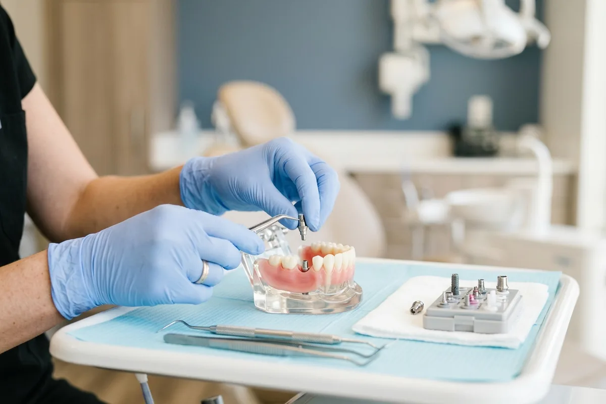
After the bone has fully integrated with the implant body, the restorative phase begins to rebuild visible tooth structure. A minor procedure may uncover the fixture to attach an abutment, which serves as the connector between the implant and the final custom prosthetic crown, bridge, or supportive dental overdenture.[1][2]
- Capturing digital or conventional impressions of the integrated implant and neighboring teeth.
- Securing a custom or prefabricated abutment connector to the underlying implant fixture.
- Fabricating the final prosthetic crown, bridge, or denture to match natural bite alignment.
- Attaching and adjusting the permanent restoration for optimal comfort, occlusion, and aesthetics.
Post-operative healing and daily maintenance
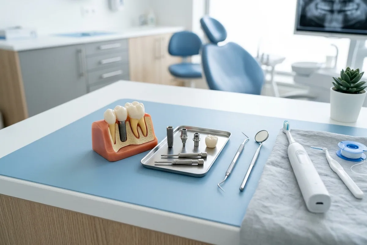
Achieving lasting success with dental implants requires diligent post-operative management and rigorous lifelong oral hygiene. While implants cannot develop dental decay, the surrounding gum tissues and underlying bone remain susceptible to biofilm-induced inflammation that requires consistent home care and professional clinical maintenance.[1][2][3]
Immediate post-operative healing expectations
Patients commonly experience mild discomfort, minor swelling, slight bruising, and intermittent oozing around the surgical site for the first 24 to 72 hours. These expected post-operative symptoms are routinely managed with conservative measures, cold compresses, and standard non-opioid pain protocols recommended by the treating clinical team.[1][2]
Daily hygiene protocols and professional surveillance
Long-term stability demands thorough daily plaque removal using soft manual or electric brushes, implant-safe floss, and specialized interdental brushes with coated wires. Patients should also maintain professional dental examinations and cleanings at least every six months so clinicians can evaluate peri-implant soft tissues and monitor crestal bone levels radiographically.[1][2][3]
Understanding complications and initiating care
Understanding potential complications empowers patients to safeguard their restorative investment through early detection and preventive habits. By partnering with a qualified dental team, individuals can address risk factors promptly and initiate tailored evaluations to restore their oral health effectively.[1][2]
Peri-implant conditions and risk reduction
Bacterial plaque accumulation can trigger peri-implant mucositis, an early reversible inflammation of the soft tissues without bone loss. If untreated, it can advance to peri-implantitis, causing progressive, irreversible bone resorption around the implant fixture. Practicing meticulous daily oral hygiene, quitting smoking, and scheduling regular professional cleanings dramatically reduce these destructive biological risks.[1][2][3]
Sources
- fda.gov — U.S. Food and Drug Administration
- mouthhealthy.org — American Dental Association
- mouthhealthy.org — American Dental Association
Common questions
Frequently asked questions
Smoking and nicotine products constrict local blood vessels in the oral tissues, significantly reducing oxygen and nutrient delivery to the surgical site. This impaired microvascular circulation hinders cellular healing, compromises bone integration, and is clinically associated with substantially higher rates of early implant failure and peri-implantitis.[1][2]
Peri-implant mucositis is a reversible inflammatory condition limited entirely to the soft gums surrounding an implant, caused by plaque accumulation. In contrast, peri-implantitis represents a more advanced, non-reversible disease state where soft-tissue inflammation is accompanied by progressive bone resorption, bleeding upon probing, and possible loss of implant stability.[1]
The biological osseointegration process typically spans several weeks to months, depending on an individual's healing capacity, systemic health, and bone quality. During this vital phase, living jawbone cells grow directly onto the implant's surface, establishing a rigid, durable anchor capable of supporting a functional prosthetic crown.[1][2]
Contact the dental team promptly if you notice persistent bleeding after applying gauze, visible discharge around the implant, or gum redness. You should also schedule a follow-up visit if the prosthetic crown or abutment screw feels loose, or if chewing produces discomfort or a bite imbalance.[1][2]
Schedule your implant consultation
Schedule a comprehensive consultation with Dr. Brian Leland, DMD to discuss your tooth replacement options and determine whether dental implants are right for your oral health needs.